
Przypadek 218. Szkoły Młodych Patologów.
Pacjent 56 letni, w trakcie diagnostyki nawracających bólów brzucha wykonano gastroskopię. W części środkowej przełyku stwierdzono szaro- żółtawego polipa wielkości do 0,5cm. Zmianę usunięto.
Do badania makroskopowego nadesłano trzy szarawe fragmenty tkankowe wym. 0,3-0,6cm.
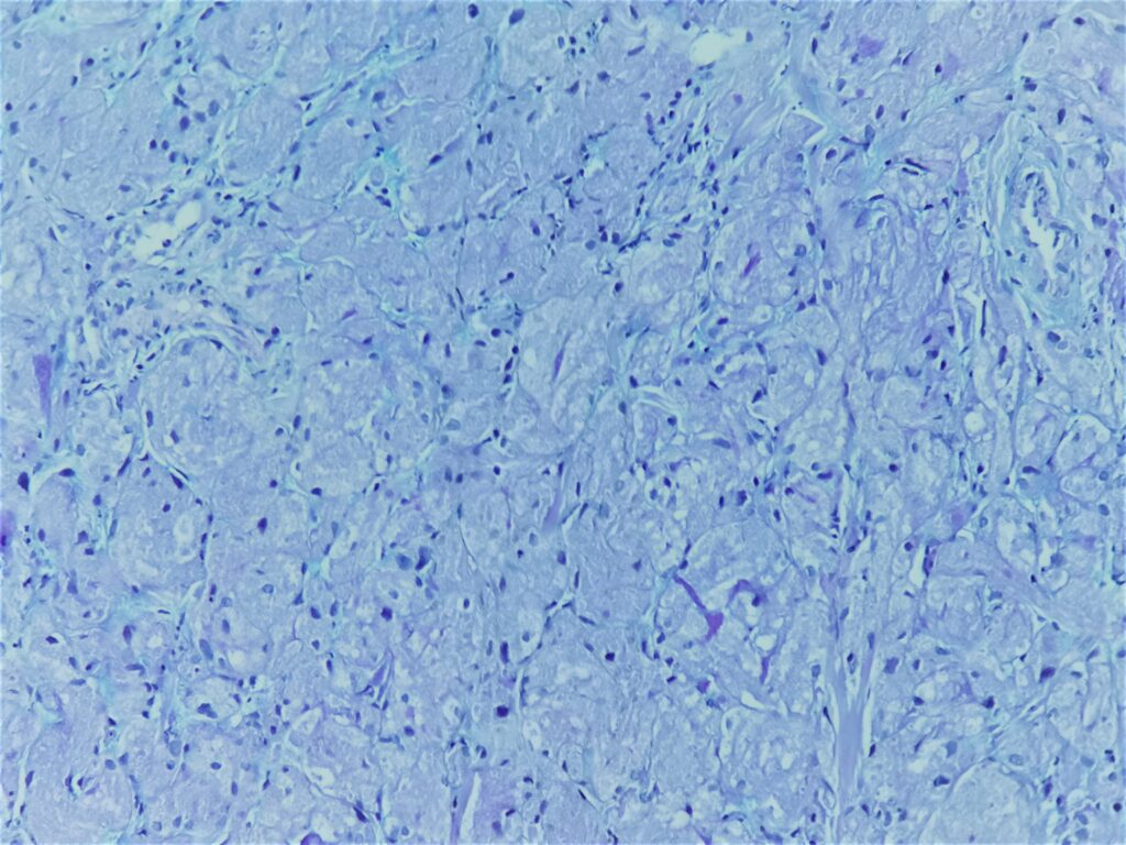
Obrazy mikroskopowe:
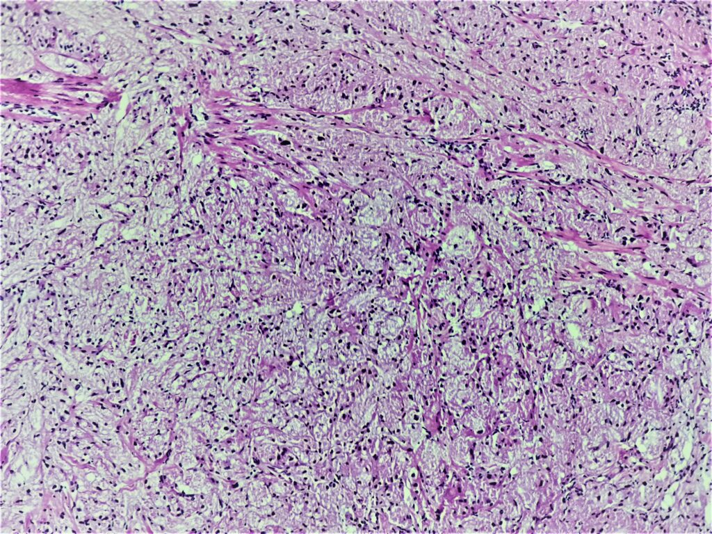

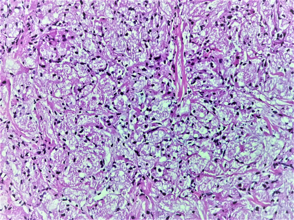
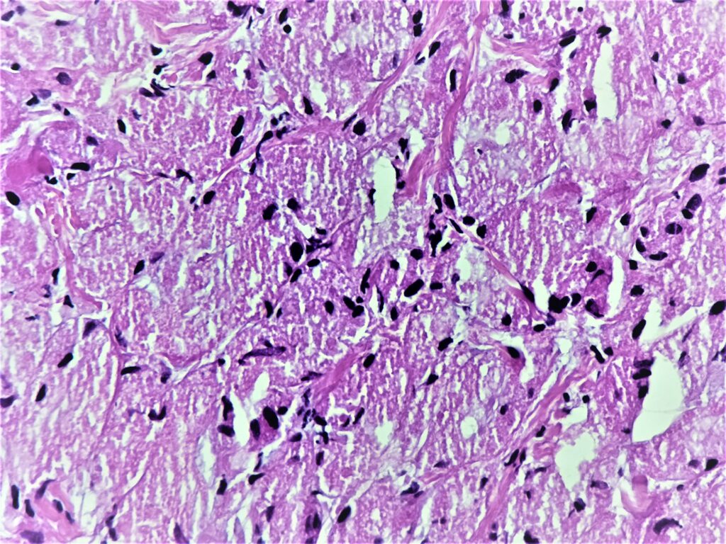
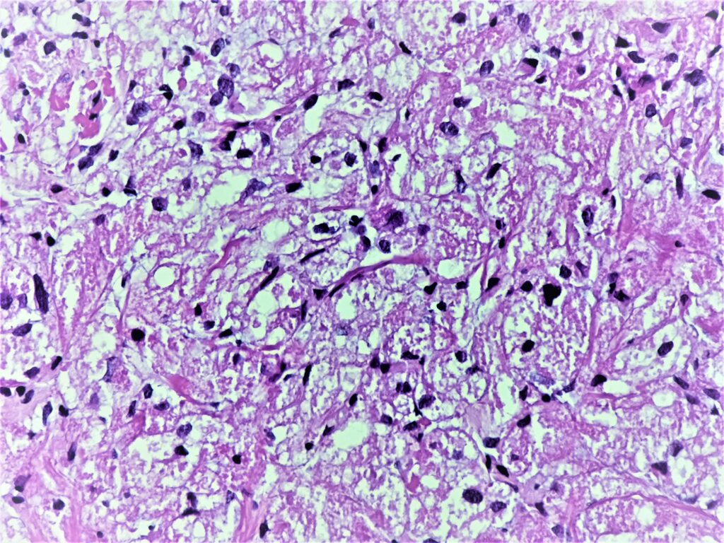
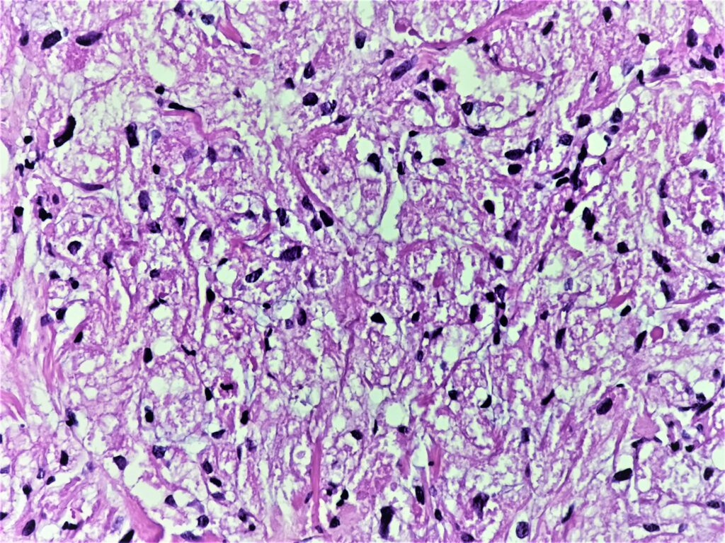

Cechy charakterystyczne:
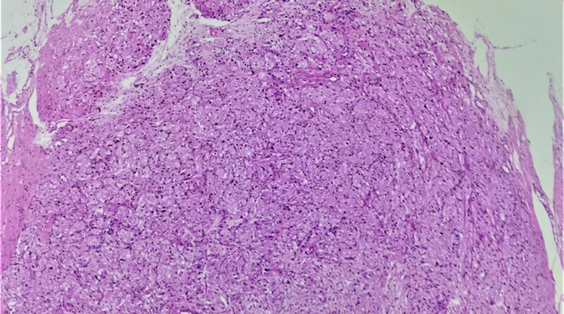
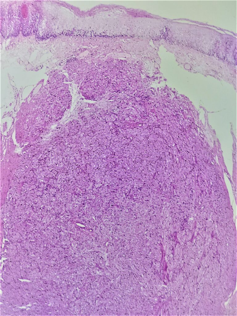
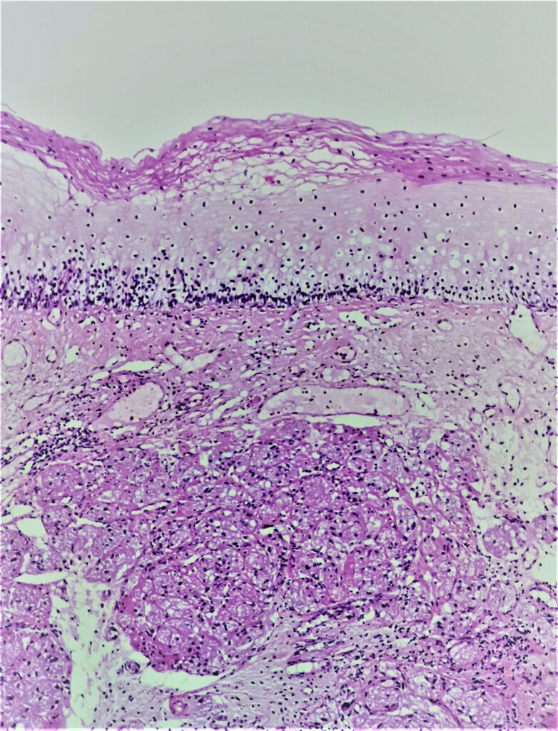
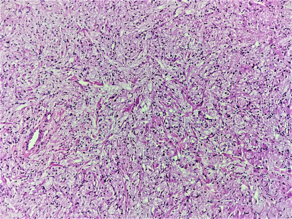
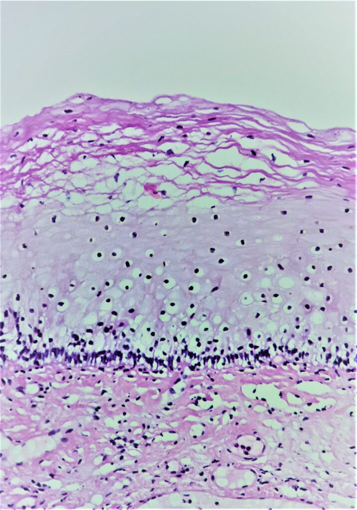
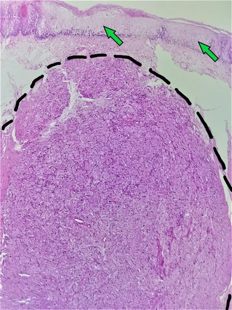
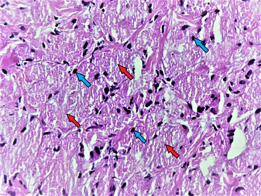
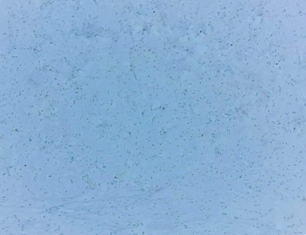
- nieotorebkowana, stosunkowo dobrze odgraniczona (czarna przerywana linia), podśluzówkowa zmiana składająca się układających się po części w drobne zespoły komórek o stosunkowo obfitej kwasochłonnej, ziarnistej cytoplazmie (czerwona strzałka), jądra komórkowe stosunkowo drobne, podobnej wielkości (niebieska strzałka), brak wzrostu stosunku jądrowo- cytoplazmatycznego, brak wyraźnych jąderek
- pokrywająca błona śluzowa zwykłego wyglądu, nabłonek wielowarstwowy płaski bez dysplazji (zielona strzałka)
- brak martwicy, licznych figur podziału, inwazji naczyniowo- limfatycznej, atypii jądrowej, wzrostu naskórkowości
Rozpoznanie:
Guz ziarnistokomórkowy przełyku (granular cell tumor of the esophagus).
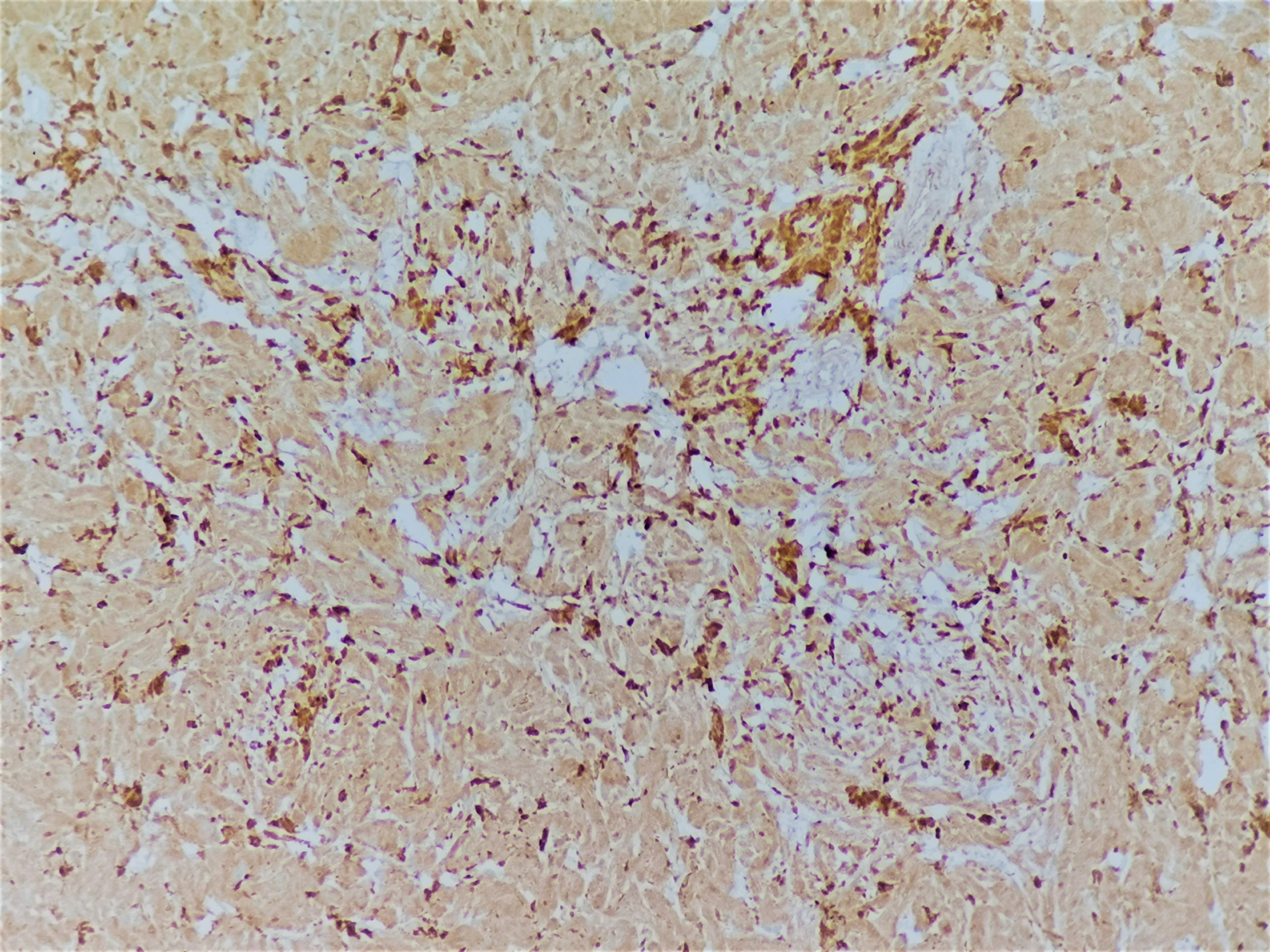
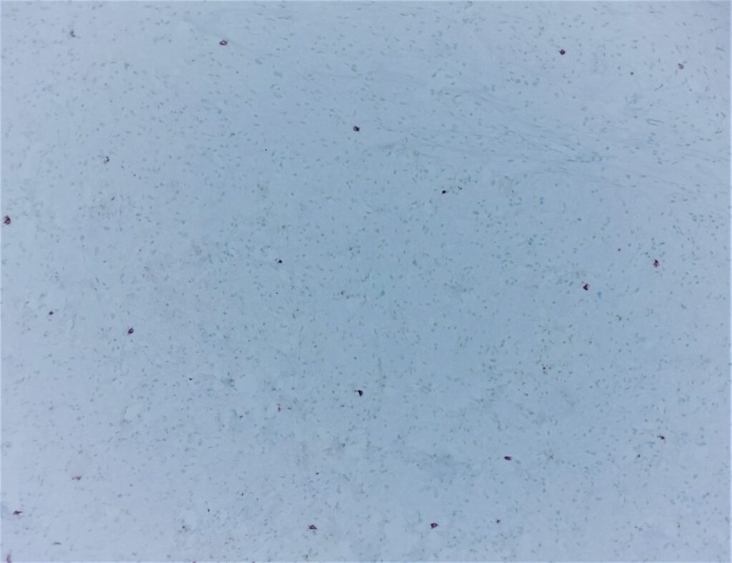
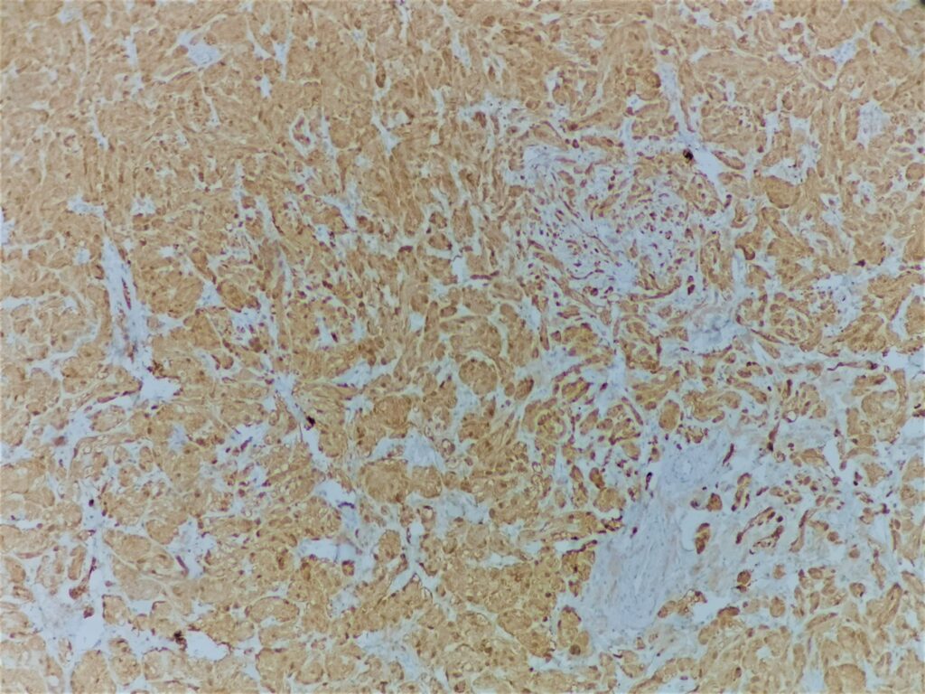
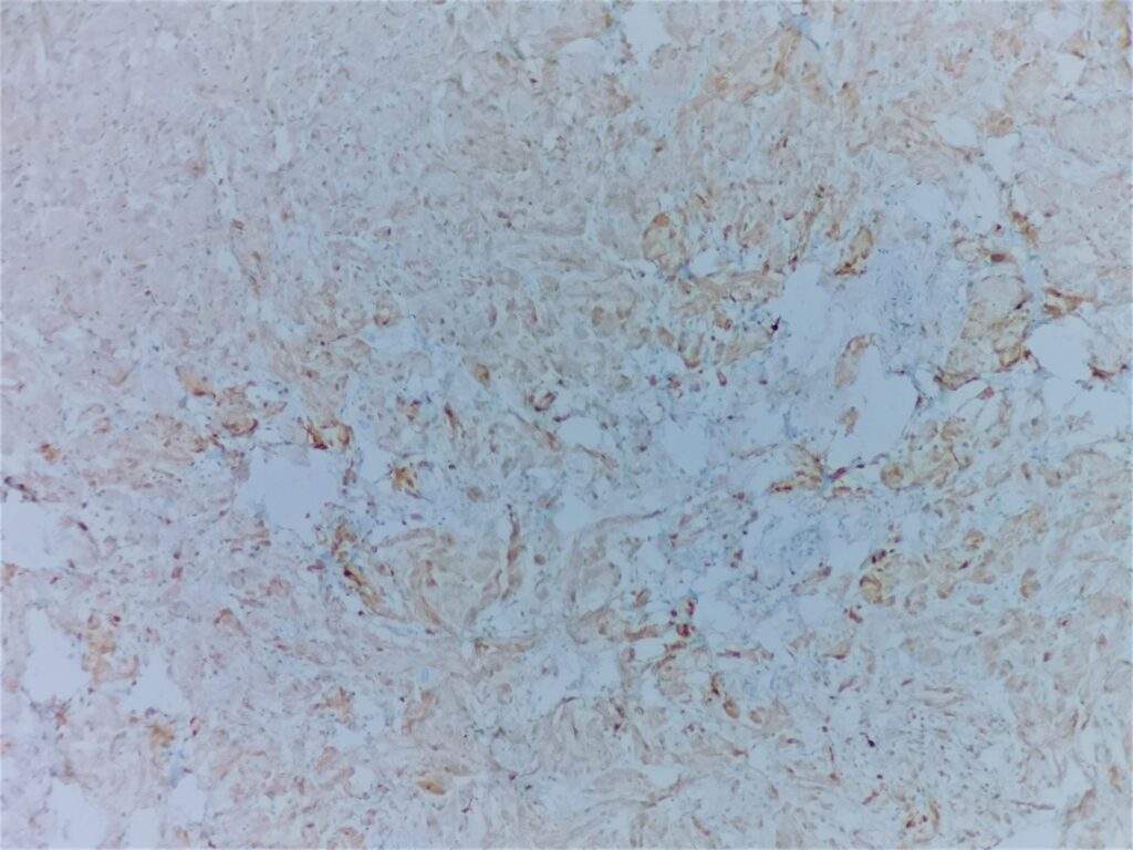
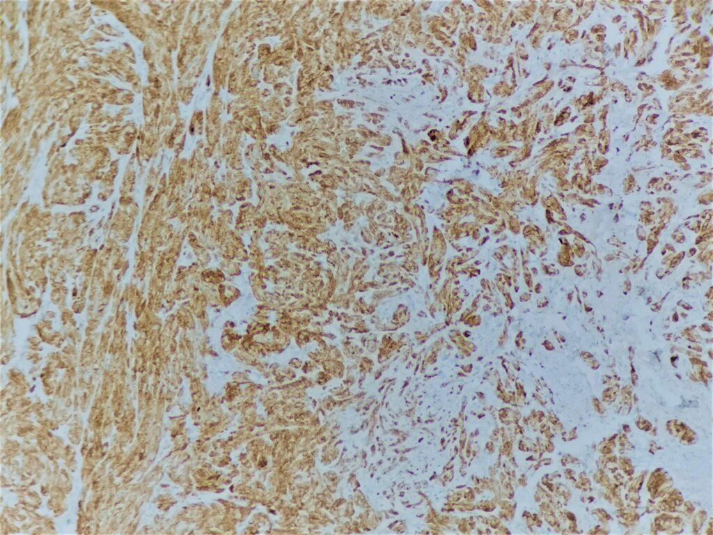
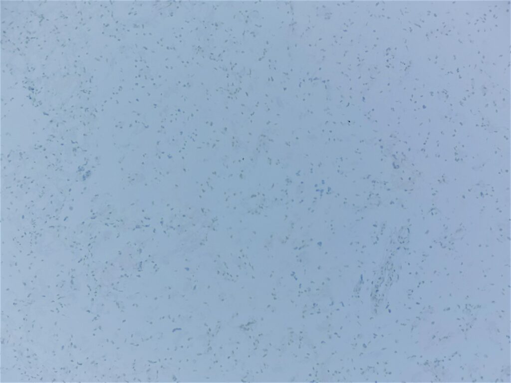
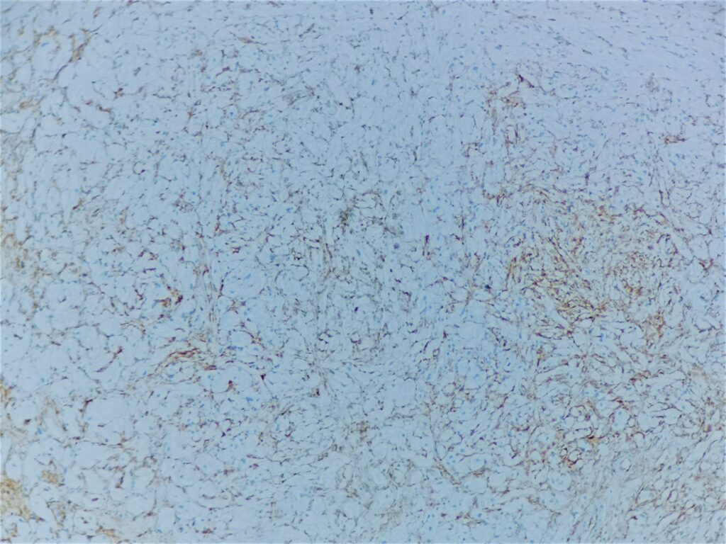
Wykonane badania dodatkowe: S100(+), CD68(+), CD117(-), CD34(-), Kalretynina (+), PanCK(-), Ki67 poniżej 1%, PAS(+).
Dodatkowe zdjęcia i informacje:
